How to Identify SNC in Your Stand: Nothophaeocryptopus gaeumannii & SNC symptoms
Proper identification of Nothophaeocryptopus gaeumannii, the fungus that causes Swiss needle cast (SNC) disease, is critical to determining whether SNC is causing the disease symptoms observed in your stand, or whether other damage agents may be to blame.
- Identification of Nothohaeocryptopus gaeumannii fungal fruiting bodies (pseudothecia)
- Symptoms of Swiss needle cast
- Nothophaeocryptopus gaeumannii life cycle
- NON-Swiss needle cast fungi & common damage agents of Douglas-fir
- Table: N. gaeumannii vs. other foliar fungi
- A closer look at N. gaeumannii with advanced microscopic techniques
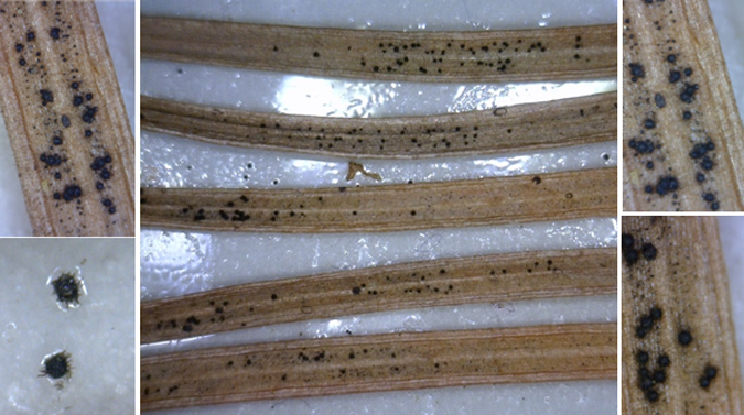
Identification of Nothophaeocryptopus gaeumannii fungal fruiting bodies (pseudothecia)
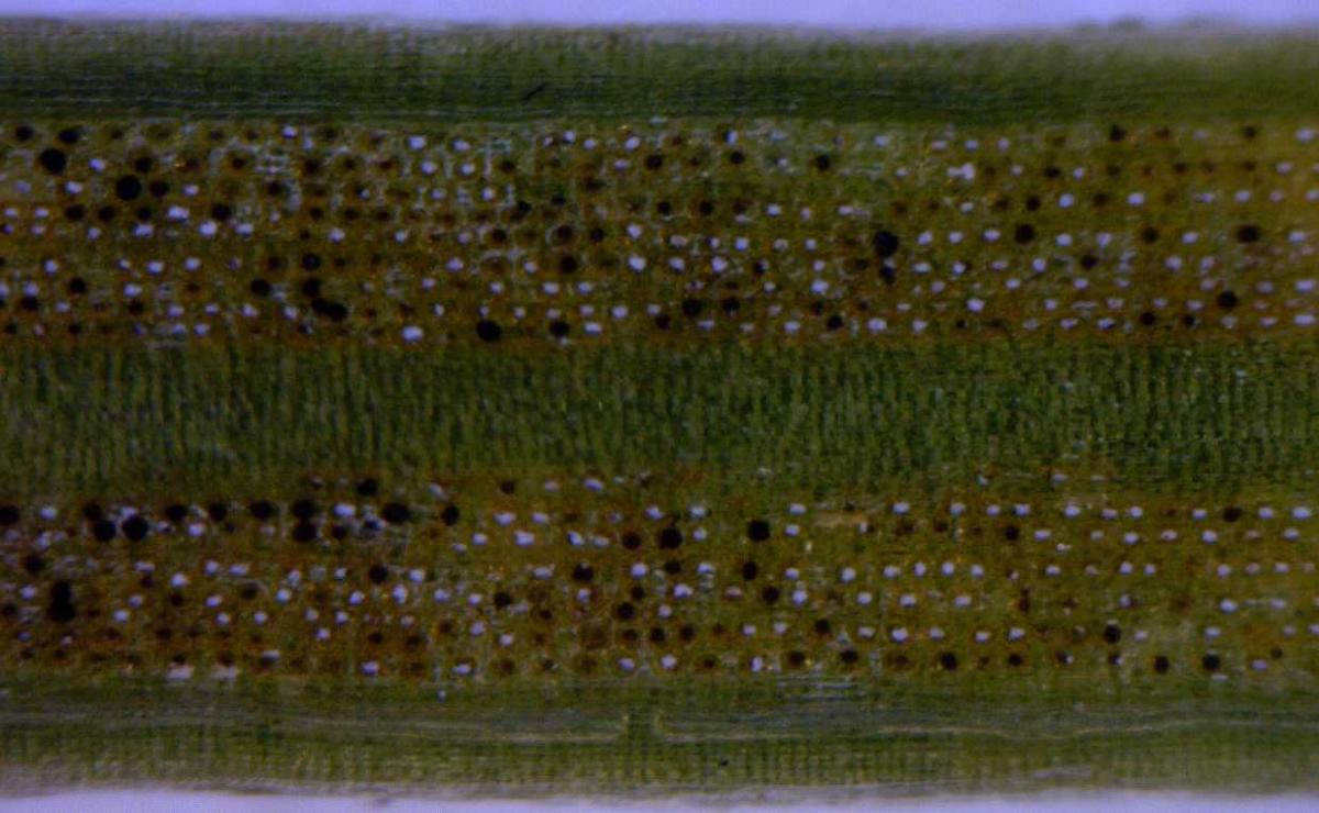
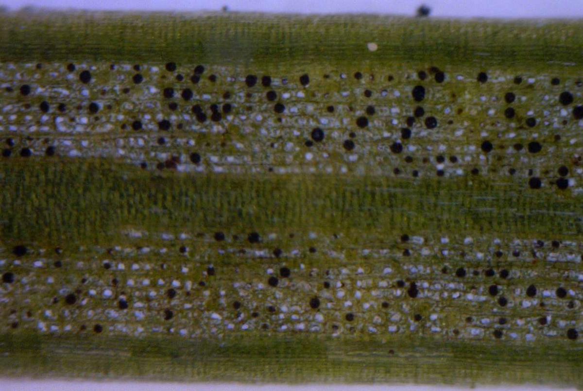
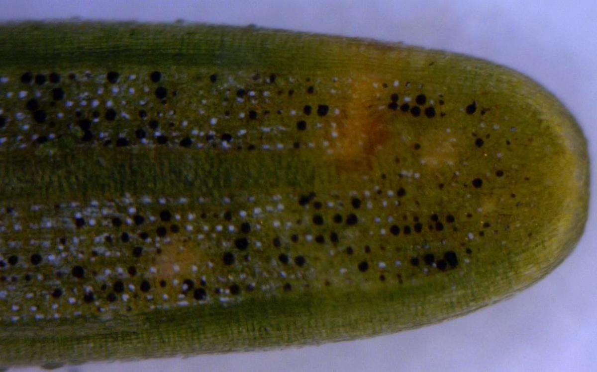
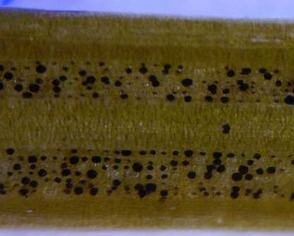
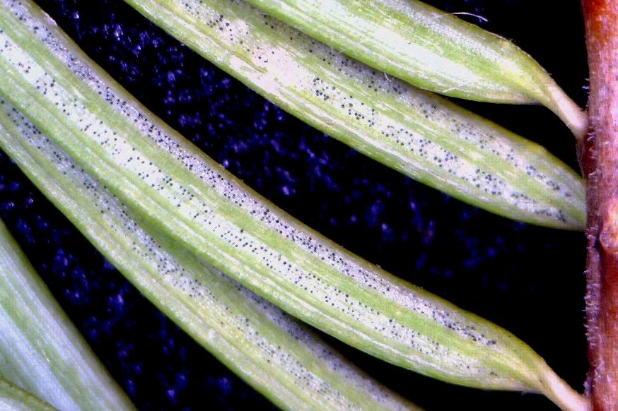
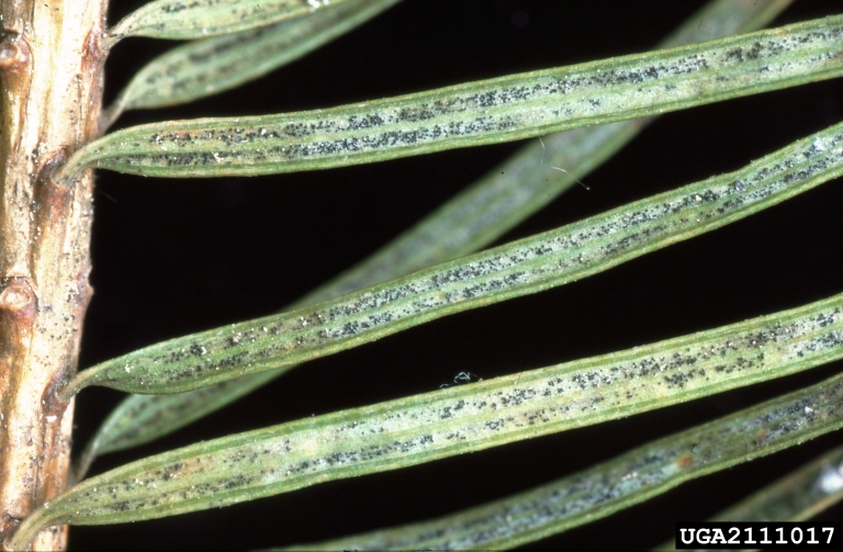
- Pseudothecia are found on the undersides of needles and occupy needle stomates
- Pseudothecia:
- are brown to black and are always centered on stomates
- have “clean” margins (not “hairy” or otherwise irregular)
- are not significantly larger than stomates (will never overlap multiple stomates)
- Pseudothecia of different sizes are frequently found on the same needle; large fruiting bodies are more mature than small fruiting bodies; larger pseudothecia are spherical, and begin to swell outward from stomates
- Proper identification of Nothophaeocryptopus gaeumannii generally requires 10x or higher magnification with a hand lens, as other fungi may look superficially similar without sufficient magnification; 40x magnification with a binocular dissecting microscope is recommended
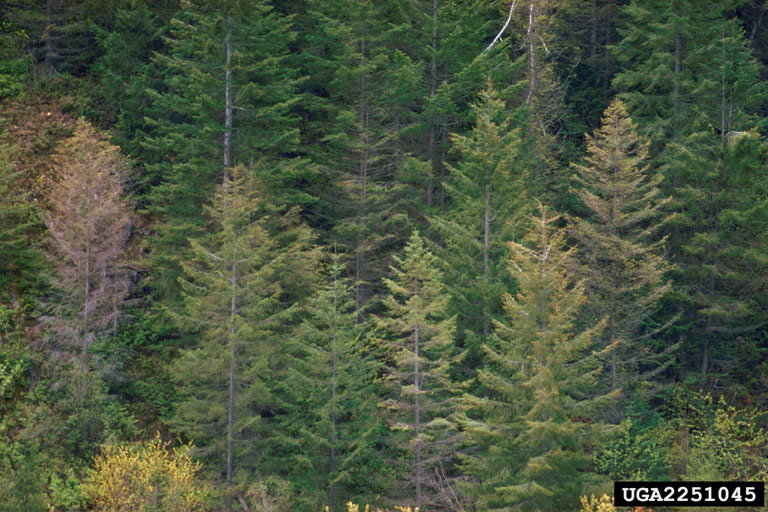
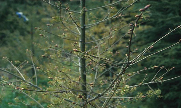
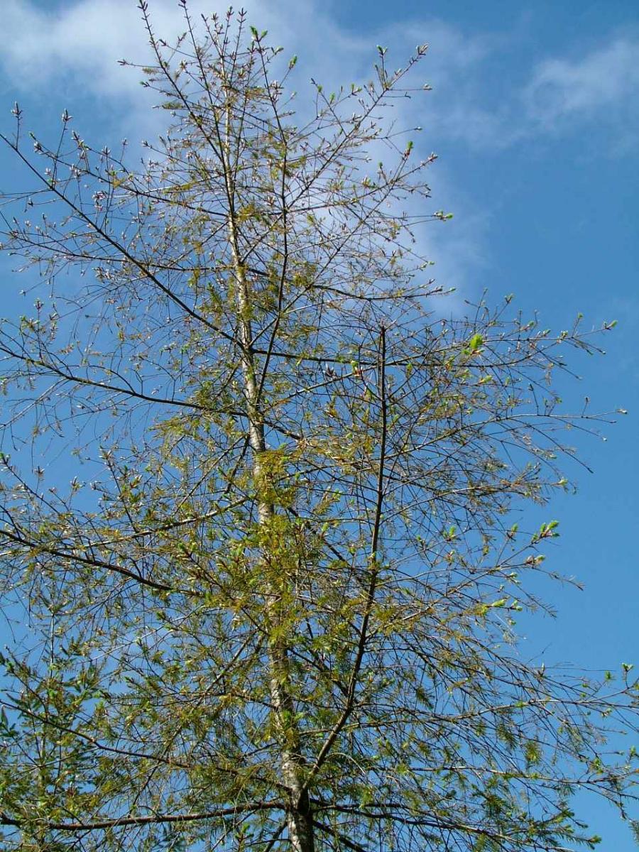
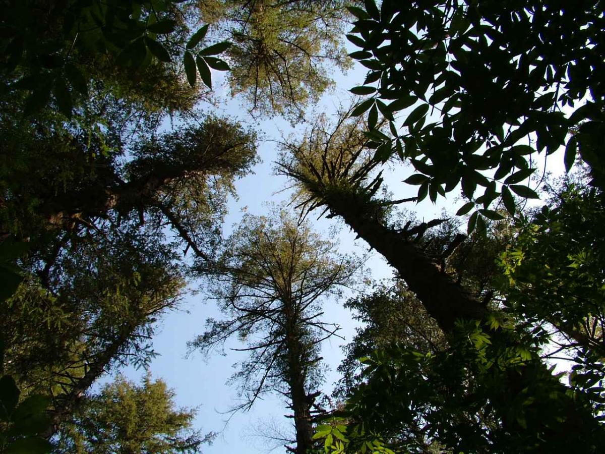
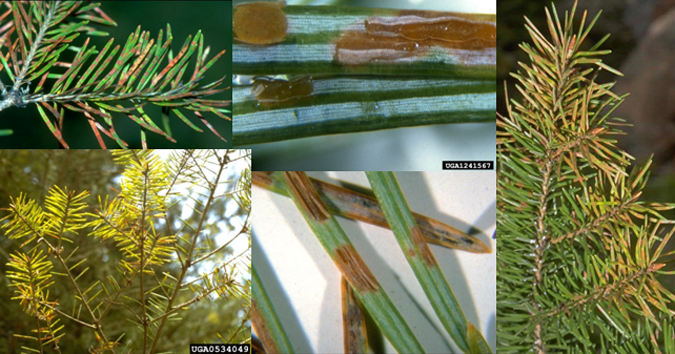
Symptoms of Swiss Needle Cast
- Chlorosis (yellow to brown coloration) of the needles and crown
- Premature needle loss (fewer than 3 years of needle retention)
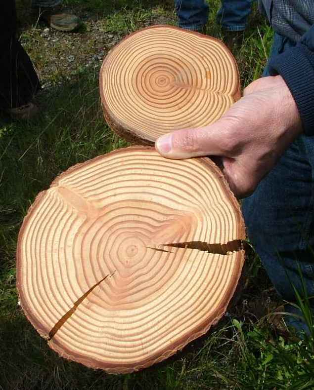
- Growth loss
- Changes to wood density and other wood properties due to increased proportion latewood
N. gaeumannii Life Cycle
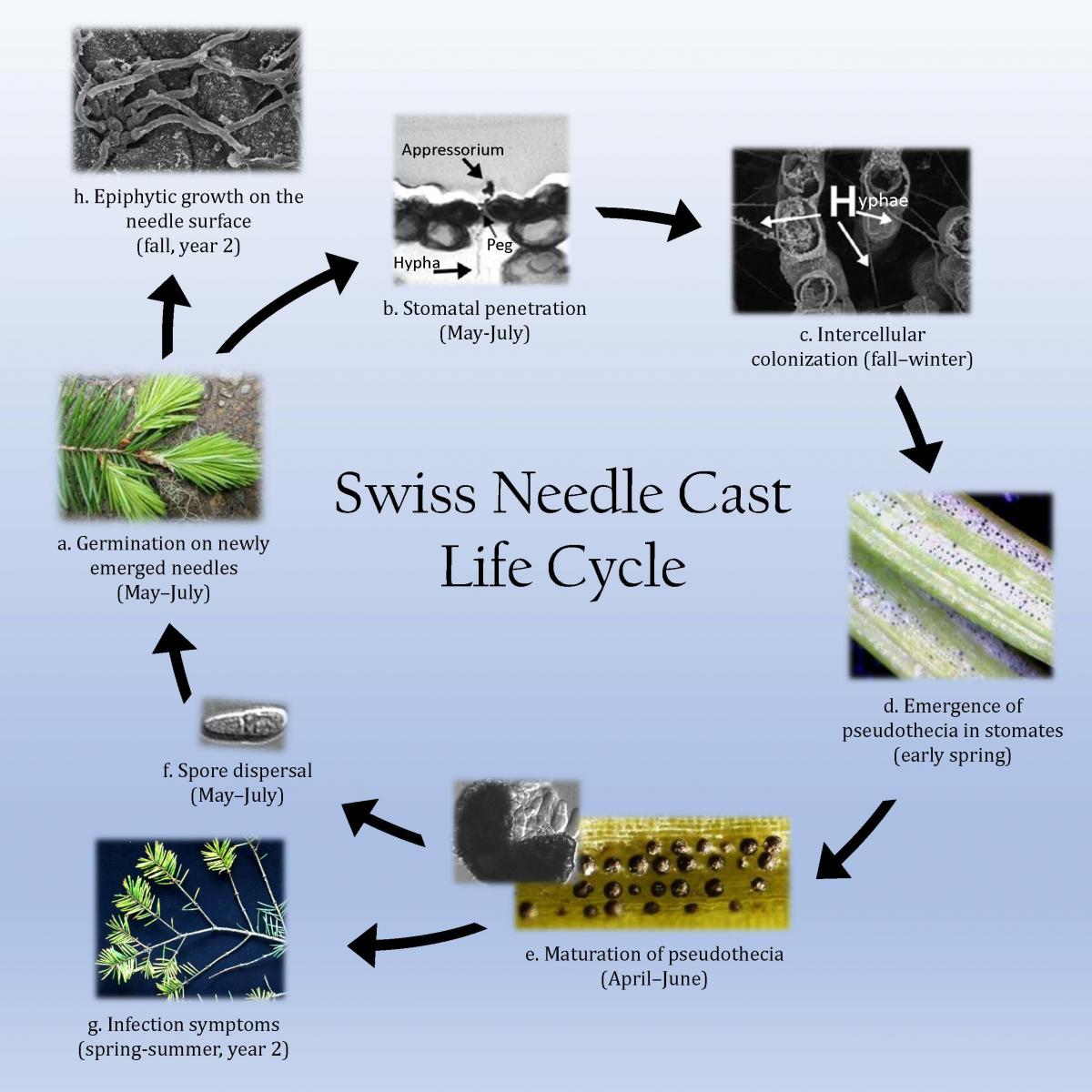
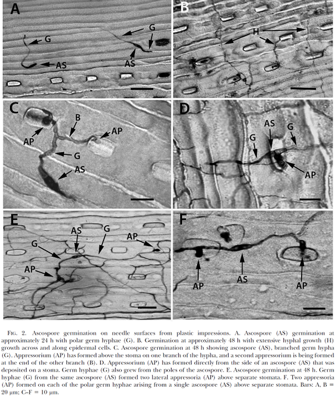
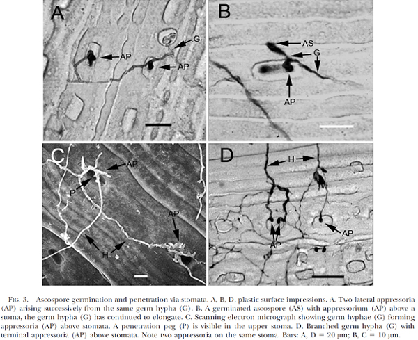
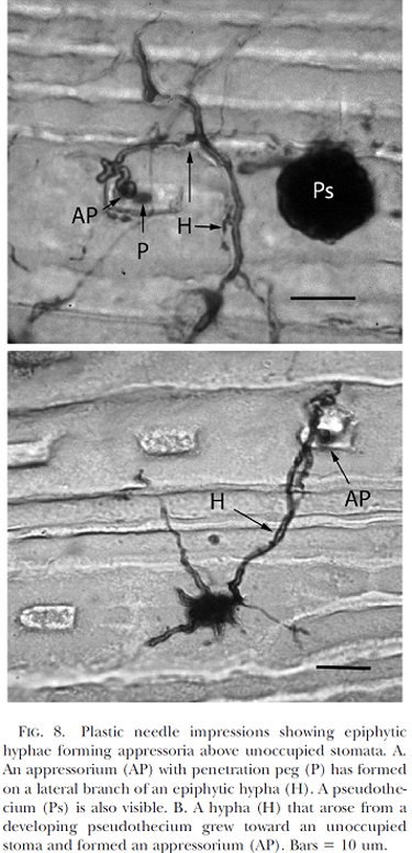
Spore dispersal during humid and wet weather begins the life cycle of Nothophaeocryptopus gaeumannii. New infections occur primarily on first-year (current year) needles and coincides with bud break and shoot elongation between May and July (a). Upon contacting a stoma, the germinating ascospore produces an appressorium from which a penetration peg is formed leading to needle penetration via the stomatal opening (b). The intercellular tissue of the needle is colonized in the fall and winter (c) and gives rise to pseudothecia, which can begin to emerge in the early spring depending on geographic location and environmental conditions (d). The fruiting bodies mature throughout the spring (e) and release ascospores between May and July (possibly into August and September) (f). Symptoms of infection can arise in second year (1-year-old) needles and older (g) and include premature needle loss (due to stomatal occlusion from pseudothecia), chlorotic foliage, and thin crowns. During the fall of the second year, epiphytic hyphae emerge from the base of pseudothecia (h) and grow along the surfaces of the needles. This epiphytic growth can result in the penetration of unoccupied stomata and might aid in expanding intercellular colonization. The development of N. gaeumannii within needles and the timing of pseudothecia development varies depending on geographic location, winter temperature, and periods of leaf wetness.
NON-Swiss Needle Cast Fungi & Common Damage Agents of Douglas-fir
- Stomiopeltis:
large brown to black spots (large enough to overlap two or more stomates); not necessarily associated with stomates; round to oval or elongated; does not protrude from needle surface

- Rasutoria:
Large, spherical, black fruiting bodies, not necessarily associated with stomates; “hairy” margins
- Rhabdocline pseudotsugae:
Rhabdocline needle cast; symptoms include red-brown banding and needle shed 12-15 months after infection; fungal fruiting in spring and fall; fruiting bodies are tan, and can be found under flaps in the epidermis on the underside of the needle, on either side of the midrib
- Cooley spruce gall adelgid (Adelges cooleyi):
Feeding on upper needles surfaces of first-year Douglas-fir needles causes yellow spots and needle kinking; insects present spring and fall; adults are covered by white, cottony wax; crawlers are small and black; damage is not usually associated with needle shed; alternate life stages for galls on spruce new growth
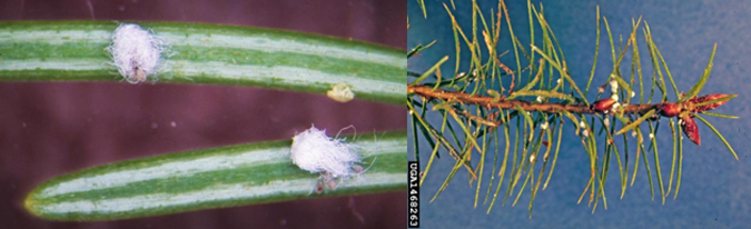
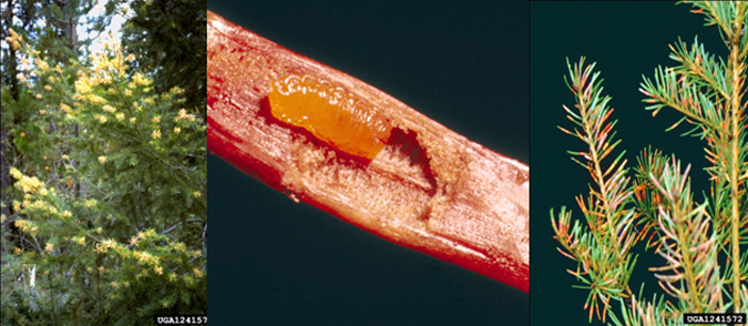
- Douglas-fir needle midge (Contarinia pseudotsugae):
Feeding by small, yellow midge larvae (legless maggots) causes yellow to red spots and swelling on new growth; insects present spring to summer and cause needle shed in late summer
N. gaeumannii vs. Other Foliar Fungi
N. gaeumannii
Pseudothecia are brown to black, are always centered on stomates, have “clean” margins, and are not significantly larger than stomates; larger pseudothecia are spherical, and begin to swell outward from stomates; fruiting bodies are present on needles year-round, and are most abundant on older needles
Stomiopeltis
Large brown to black needle spots (large enough to overlap multiple stomates); not necessarily associated with stomates; round to oval or elongated; does not protrude from stomates; grows superficially (on needle surface) and does not cause significant damage or needle shed
Rasutoria
Large black fruiting bodies (often large enough to overlap multiple stomates), often associated with stomates; “hairy” margins; protrude from stomates; only weakly parasitic and rarely cause needle senescence or shedding
Rhabdocline
Tan fruiting bodies on the undersides of needles beneath flaps in the epidermis; similar needle shed symptoms to SNC, but red brown needle banding and distinctive fruitin
For more images of damage agents of Douglas-fir, follow these links to forestryimages.org:
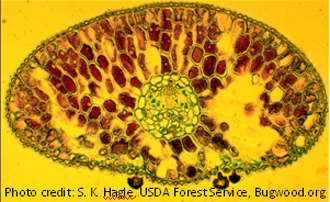
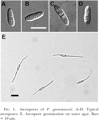
A closer look at N. gaeumannii with advanced microscopic techniques
- Cross section of a pseudothecium
- Ascospores and germinating ascospores on water agar
- Germinating ascospores on needle surfaces
- Ascospore germination and penetration via stomata
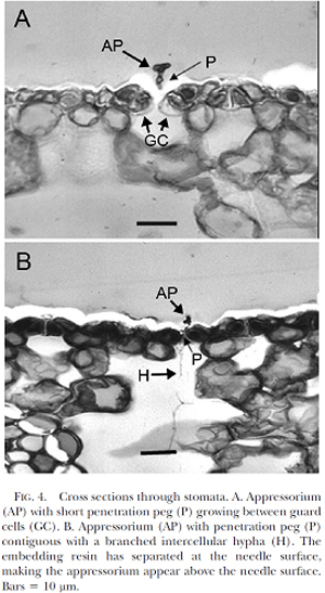
- Cross sections through stomata
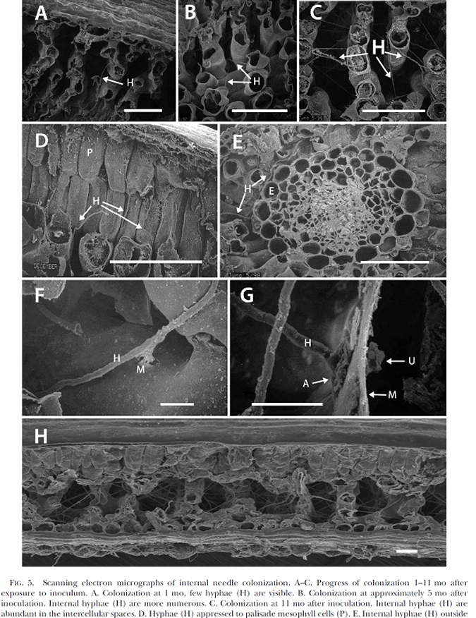
- Scanning electron micrographs of internal needle colonization
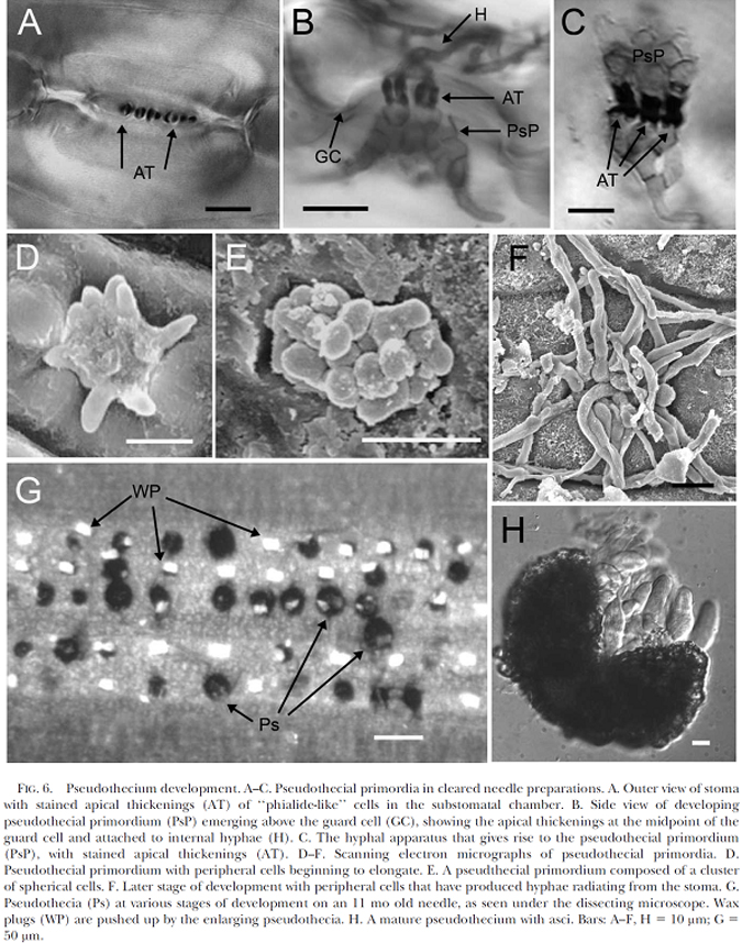
- Pseudothecium development
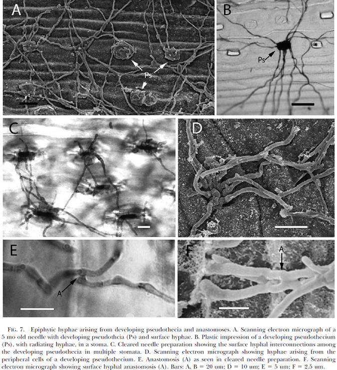
- Epiphytic hyphae